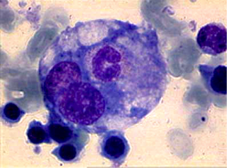
An example of hemophagocytosis.
Salmonella enterica is a bacterial pathogen notorious for causing food poisoning symptoms that are uncomfortable at best and deadly at worst. Infamously linked to raw eggs, it is the reason many of us were cruelly prevented from licking cake batter right off the spoon when we were younger. Many will also recognize S. enterica for its role in typhoid fever, an infection that is most prevalent in the developing world where sanitation is limited.
Strains of S. enterica can be further classified into serotypes, or distinct variations of bacteria, based on the molecules on their cell surfaces. Some serotypes can cause more serious illness than others—that’s why some strains of S. enterica cause mild discomfort and some can be fatal. Typhoid fever is caused by the serotype of S. enterica Typhi or Paratyphi, and results in mild to severe flu-like symptoms. It can be deadly, but it is also possible to carry the bacteria and have no symptoms at all, facilitating the spread of this debilitating disease.
Dr. Corrie Detweiler and her lab in the department of Molecular, Cellular and Developmental Biology study Salmonella enterica in the context of the host cells that harbor S. enterica—macrophages. Macrophages are white blood cells of the immune system that police our bodies in search of pathogens such as bacteria or viruses. Upon encountering a pathogen, the macrophage will attempt to phagocytose, or engulf, it and subsequently destroy it. To successfully infect humans, S. entericacolonizes macrophages in order to avoid being targeted for phagocytosis. Think of a spy infiltrating a city under siege—he or she will hide in a house rather than risk being recognized on the streets.
When studying how the bacteria infect host cells, the Detweiler lab uses different serotypes ofS. enterica. In addition to Typhi, the lab uses the serotype Typhimurium, a strain of the bacteria that causes gastroenteritis (infectious diarrhea) in humans and other mammals. Importantly for the Detweiler lab, mice exhibit typhoid-like symptoms when infected with S. Typhimurium, making this system an ideal model for the study of typhoid fever.
Hemophagocytes (HMs) are a specific type of macrophage that have engulfed erythrocytes (i.e. red blood cells) and leukocytes (i.e. white blood cells). This vampiric process of “blood eating” is called hemophagocytosis and is considered a state of disease because the blood cells are intact and healthy but are somehow targeted for engulfment by macrophages. HMs are often seen as a response to severe infection by bacteria, viruses and parasites. It was previously reported that S. Typhimurium resides within HMs, specifically within HMs containing leukocytes (known as leukophagocytes). But the Detweiler lab found, by fluorescence microscopy, that the bacteria reside in HMs containing erythrocytes (known as erythrophagocytes) as well as leukophagocytes.
Dr. Carolina Pilonieta, a former postdoc in the Detweiler Lab and leader of this project, was especially excited by this finding:
“It hadn’t been seen before because it was really hard to test. This was the first time anyone had created a model for studying HMs in vitro and gotten it to work efficiently. Now we’ve opened the doors to allow us to exploit HMs in future work.”
Carolina wanted to know whether they could induce erythrophagocytosis (engulfment of red blood cells) by treating cells with interferon gamma (IFNγ; a signaling molecule produced by immune cells in the presence of an invading pathogen) and lipopolysaccharide (LPS; a component of the surface of bacteria). Both of these induce an immune response in mouse cells. If erythrophagocytosis was induced, this would strengthen the argument that the process of red blood cell engulfment is a response to bacterial invasion and not just an artifact of keeping cells in dishes.
The researchers used both IFNγ and LPS to stimulate hemophagocytosis in mouse macrophages. They then lysed, or broke open, all erythrocytes left uneaten. Any erythrocytes detected after this treatment were assumed to have been engulfed by the macrophages. The hypothesis that IFNγ and LPS treatment could cause erythrophagocytosis proved correct—engulfment of red blood cells was significantly increased in the mouse macrophages.
In her next experiments, Carolina wanted to confirm that exposure to S. Typhimurium cells stimulates erythrophagocytosis. She found that S. Typhimurium strongly increases erythrophagocytosis, and her initial conclusions were that this response is specific toSalmonella. However, more recent studies in the Detweiler lab have suggested that it may instead be a general response to bacterial invasion—work that Carolina hopes will be published in the next year.
In her own words: “I liked the story better as Salmonella causing it… but you have to follow where the data goes.”
Carolina went on to show that S. Typhimurium cannot force macrophages to eat non-reactive, inert polysterene beads. This suggests that something (a cell-surface protein, for example) on the erythrocytes themselves is being recognized by the macrophages, making the engulfment specific. Macrophages that had engulfed erythrocytes were much more likely to be infected by S. Typhimurium, and the lab is hypothesizing that the bacteria is manipulating the macrophage to erythrophagocytose (i.e. engulf erythrocytes) —providing a survival niche for them to proliferate.
S. Typhimurium does not infect red blood cells, nor does it require red blood cells to be eaten by a macrophage before it can colonize that macrophage. So what could be the advantage of residing in erythrophagocytes? HMs degrade heme, which is a molecule that stores oxygen in the blood. Heme is in high concentration in red blood cells, and contains iron, so erythrophagocytes could have a higher abundance of this necessary metal than other cells. The degradation of heme could also play a role in making a less toxic environment for bacteria within HMs—the degradation process interferes with white blood cells’ ability to neutralize invaders.
Think again of that spy in an enemy city—he or she can infiltrate a house, take the inhabitants hostage, and then force the hostages to order in supplies. Order the right pizza, for example “one red blood cell special, extra-large,” and not only do you feed yourself—you keep your hostages happy and less likely to kill you!
It is tempting to ask why the bacteria does this, because it seems like a relatively complicated way to acquire more nutrients and protect itself from the host immune response. But a “Why?” question implies that the bacteria is fully in control of its own evolution, and that any adaptations in host-pathogen interactions are made with a greater plan in mind. Instead, we should think of this process as something that came about accidentally, by an accumulation of small changes, each of which were advantageous to the bacteria’s survival. The final result of many years of evolution in S. Typhimurium is a neat trick of manipulation that creates a protected enclave in which the bacteria can survive, while the host’s defense system remains waiting and watching outside.
By Alison Gilchrist
